cellXpress 2.4.0 with WSI support available now (June 2024)
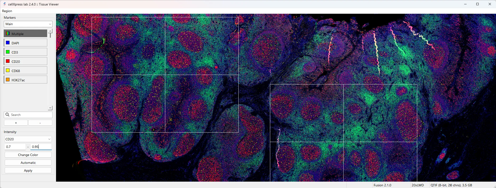
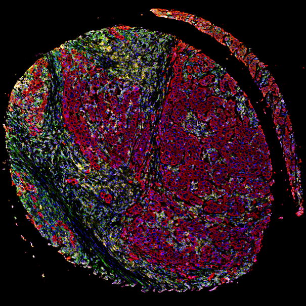
Visualize and analyze huge whole-slide tissue images
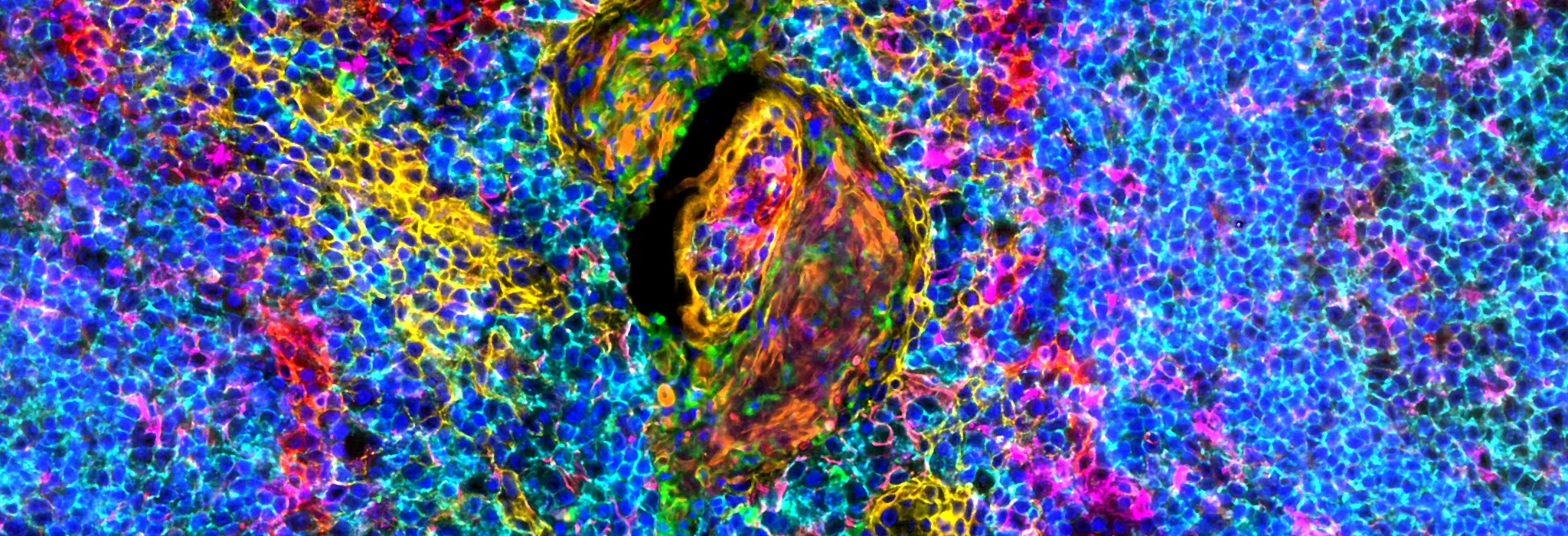
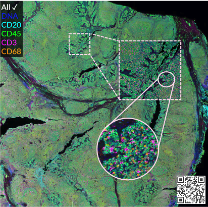
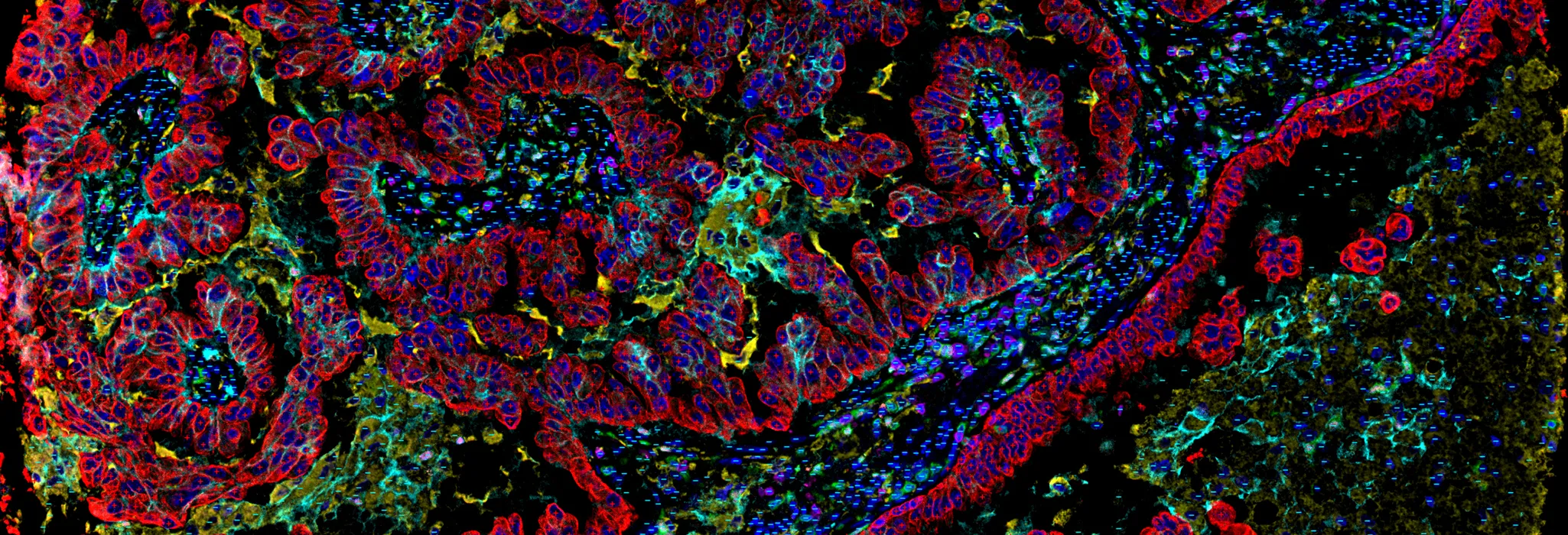
Visualize highly-multiplexed cellular and tissue images
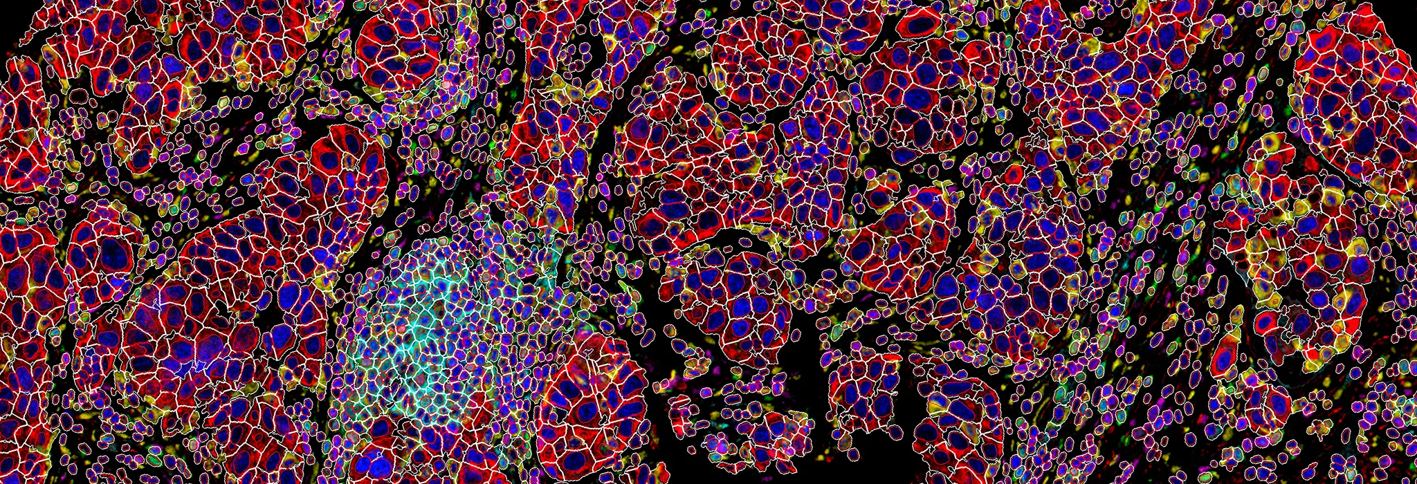
Detect single cells in tissues using CellShape AI
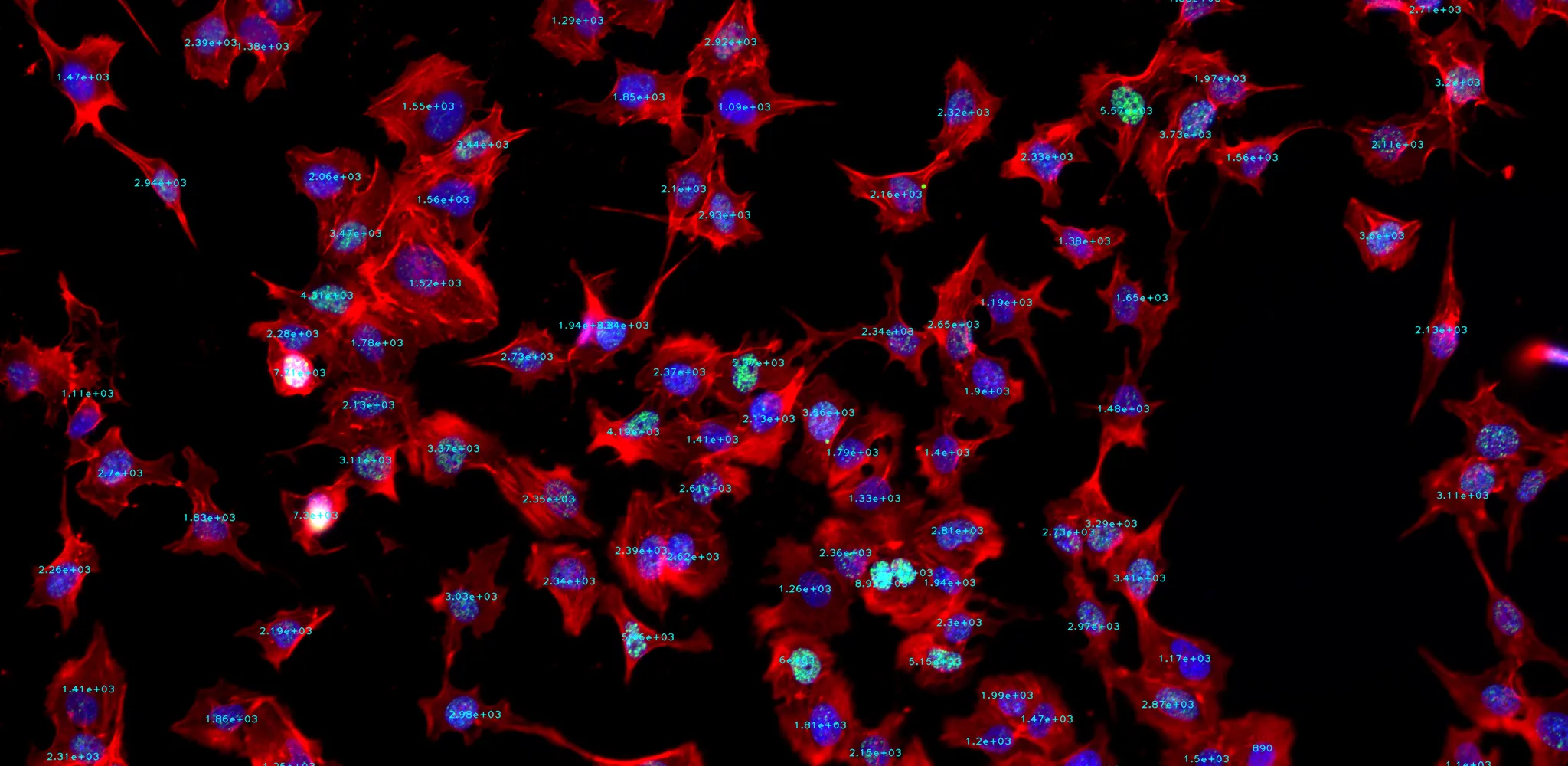
Quantify and visualize single-cell and subpopulation phenotypes
Discover spatial organizations of disease-relevant cellular subpopulations
Key features
Marker-set architecture

Optimum image processing based on different marker sets
Hyperplexed images

Intuitive visualization of large tissue images and results with 50+ markers
CellShape AI segmentation
Accurate detection of heterogenous and overlapped cells in tissues
Subpopulation identification

Systematic identification of diverse cell types or subpopulations
Published work that used cellXpress
Choice of PD-L1 immunohistochemistry assay influences clinical eligibility for gastric cancer immunotherapy
J Yeong et. al., Gastric Cancer, 2022
cellXpress was used to quantify the three PD-L1 antibody clones (22C3, SP142 and 28-8) and cytokeratin.